ROIs
ROI masks are provided for all subjects and cover retinotopically defined visual areas, category-selective regions from functional localizers, motion-selective areas, and visual-stream parcels. The ROIs are provided as a suggested common reference for downstream analyses; the underlying contrasts and retinotopic maps will be released too to enable alternative definitions.
Available ROI Sets
Set |
ROIs |
Source |
Criterion |
|---|---|---|---|
Retinotopic |
V1v, V1d, V2v, V2d, V3v, V3d, V3A, V3B, hV4, LO1, LO2, TO1, TO2, VO1, VO2, IPS0, SPCS, IPCS, EVC |
Retinotopy localizer |
Polar angle reversals, eccentricity gradients (R²-thresholded) |
Face-selective |
OFA, FFA-1, FFA-2, pSTS-faces |
fLoc |
faces > others |
Body-selective |
EBA, FBA |
fLoc |
bodies > others |
Scene-selective |
PPA, OPA, MPA, SPL |
fLoc |
scenes > others |
Character-selective |
VWFA-1, VWFA-2, mfs-words |
fLoc |
characters > others |
Object-selective |
l-objects, v-objects |
Object localizer |
objects > scrambled |
Motion |
MT, MST |
Motion localizer |
motion > static, ipsilateral > static |
General visual selectivity |
laion-general |
LAION main experiment |
GLMsingle R² |
Sectors of visual selectivity |
laion-EVC, laion-dorsal, laion-lateral, laion-ventral |
Retinotopic ROIs + anatomical borders |
Subdivision of laion-general |
Functional ROIs
Design Choices
All functional ROIs were manually delineated on the cortical surface. Statistical maps informing the delineations, such as polar angle, or localizer contrast t-statistics, were rendered as flatmaps via pycortex [Gao2015] and annotated in Inkscape.
Selection of ROIs. ROIs were included based on relevance to the field and consistent identifiability across subjects.
Mutual exclusivity. ROIs can overlap across sets but not within a set. For example, face-selective FFA-1 may overlap with body-selective FBA, but not with face-selective FFA-2.
Contrast thresholds. Contrast thresholds used in the definitions of ROIs vary across the field and are dependent on the statistical power of the experiment. To accommodate different user preferences, we applied a liberal threshold at t>0 and relied on human judgment to identify steep drop-offs in functional preference. The resulting ROIs may be larger than those reported in studies applying more conservative thresholds. We will soon release the underlying t-statistic maps, allowing users to apply stricter thresholds within the provided ROIs as their analyses require.
Spurious activity in early visual cortex. Some category localizer contrasts yield significant responses in V1–V3, likely reflecting low-level stimulus confounds rather than genuine category selectivity. Such activations are excluded from all category-selective ROI labels.
Retinotopy-derived ROIs
Boundaries are traced on pRF parameter maps.
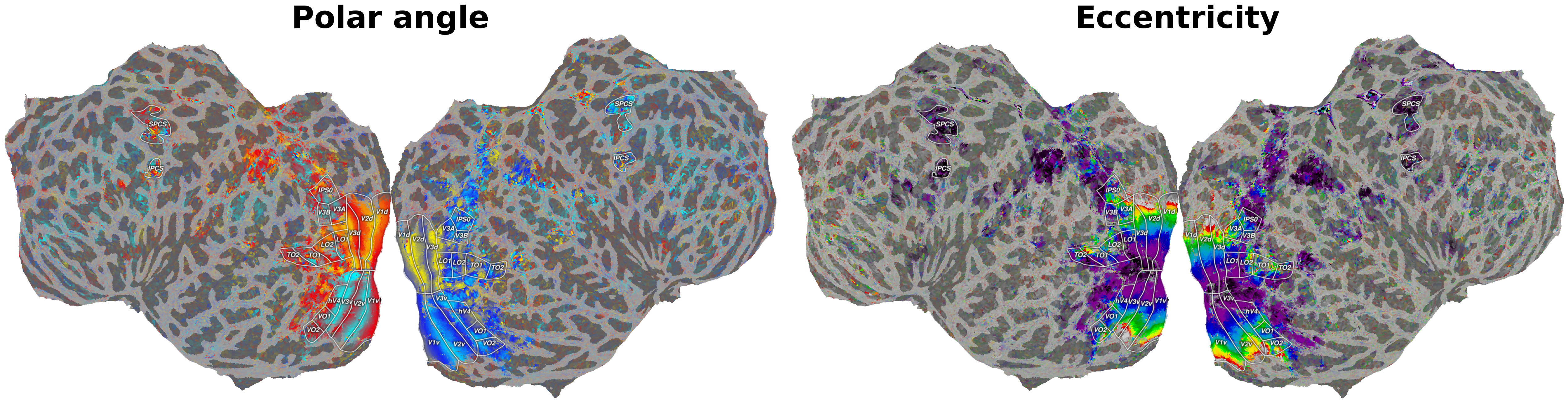
Retinotopy-derived visual area ROIs.
V1 / V2 / V3 are defined by successive polar-angle reversals and
released as separate dorsal (*d, lower visual field) and ventral (*v, upper
visual field) ROIs. EVC is the union of V1-V3, provided as a convenience mask for
analyses targeting early visual cortex as a whole.
Note
EVC is not to be confused with laion-EVC. laion-EVC is restricted to vertices showing significant responses in the LAION experiment and is typically smaller than the retinotopically defined EVC.
V3A / V3B lie anterior to V3d near the transverse occipital sulcus. They share a foveal representation; V3A on its medial side and V3B on its lateral side.
IPS0 extends along the intraparietal sulcus from V3A/V3B, with a polar angle progression from the upper vertical meridian (UVM) to the lower vertical meridian (LVM).
hV4 sits lateral to V3v. The anterior boundary is an eccentricity reversal. hV4 contains a full hemifield representation.
VO1 / VO2 are anterior to hV4 on the ventral surface. They share another foveal cluster and can be separated by a UVM representation.
LO1 / LO2 / TO1 / TO2 cover the lateral occipitotemporal cortex. LO1 extends laterally from V3d. Each area contains a hemifield representation and their boundaries are defined by polar angle reversals. LO1 and LO2 share the foveal confluence of EVC whereas TO1 and TO2 contain a separate foveal representation.
SPCS / IPCS (superior and inferior precentral sulcus) are defined by binarizing the population receptive field R² map without polar angle delineation; only clear clusters on the precentral sulcus are labeled.
Localizer-derived ROIs
Localizer-derived ROIs are defined from the fLoc and motion localizer experiments. For details on the localizer paradigms, see Functional Localizers.
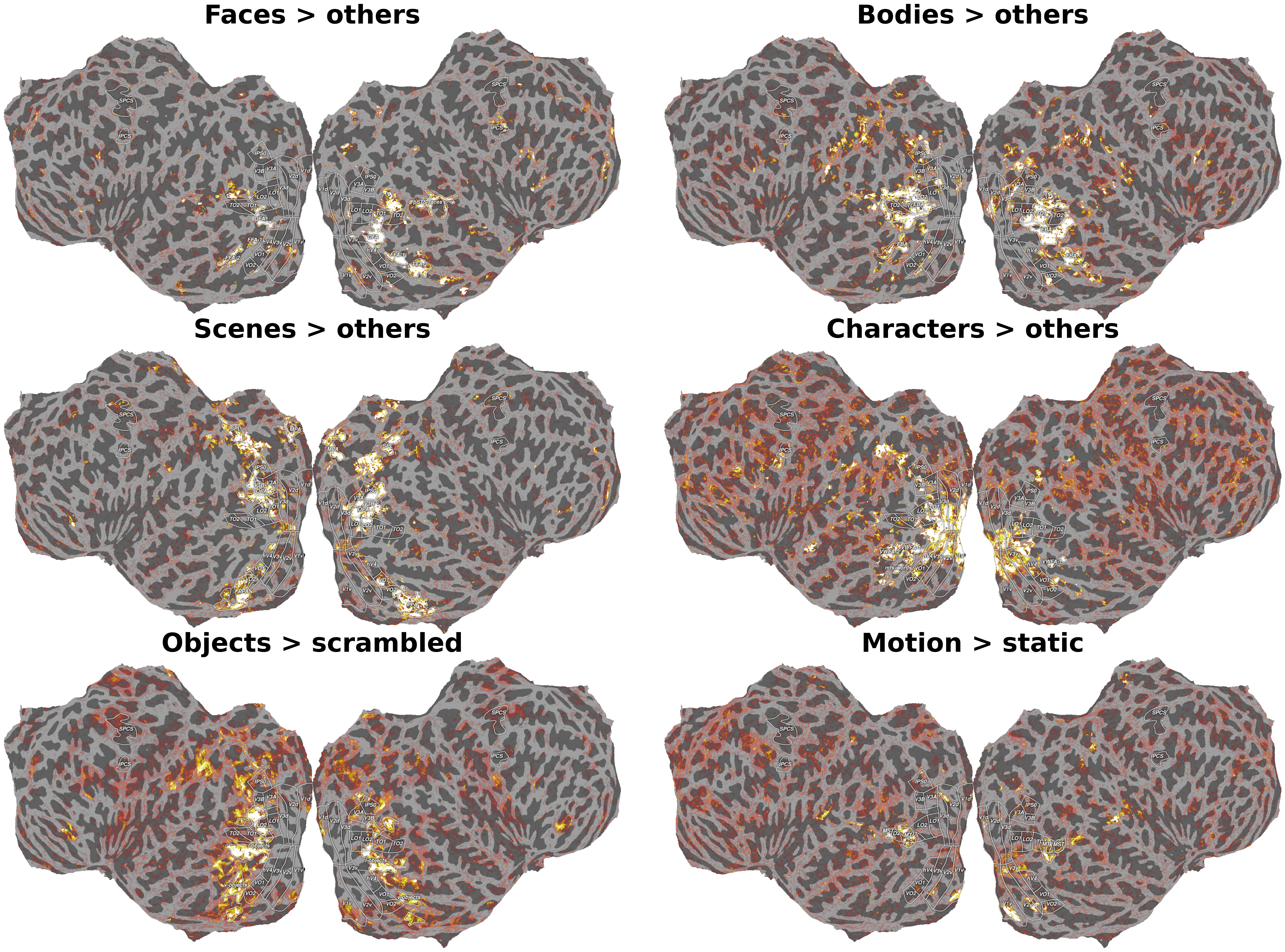
Localizer-derived category-selective ROIs.
Face-selective (faces > others). OFA (IOG-faces) is on the inferior occipital gyrus, lateral to hV4. FFA-1 (posterior fusiform, pFus-faces) and FFA-2 (mid-fusiform, mFus-faces) are on the fusiform gyrus: FFA-1 sits posteriorly on the lateral fusiform gyrus; FFA-2 is a separable cluster anterior to FFA-1. pSTS-faces is on the posterior superior temporal sulcus (pSTS), anterior to EBA, and kept separate from EBA.
Body-selective (bodies > others). EBA is a large complex on the lateral occipitotemporal cortex surrounding motion-selective MT/MST. FBA is on the fusiform gyrus lying approximately on the boundary of FFA-1 and FFA-2.
Scene-selective (scenes > others). PPA is a contiguous cluster of selectivity along the collateral sulcus. Separated clusters in VO1 are not included. OPA lies around the transverse occipital sulcus. MPA (also known as retrosplenial cortex, RSC) is in medial parietal cortex. We tentatively delineate a fourth scene-selective area, SPL, on the superior parietal lobule, dorsal and anterior to OPA. Bridging activations connecting MPA and OPA are discarded.
Character-selective (characters > others). VWFA-1 is the first contiguous character-selective region in the posterior occipitotemporal sulcus (pOTS) and surrounding banks. VWFA-2 is the second character-selective region on the mid-occipitotemporal sulcus (mOTS). Clusters of character-selective activation entirely outside the occipitotemporal sulcus (OTS) are not labeled as VWFA. mfs-words is a mid-fusiform cluster distinct from the OTS patches, labeled where identifiable.
Object-selective (objects > scrambled). Object selectivity is subdivided into two mutually exclusive sectors using a coarse lateral/ventral anatomical boundary: the inferior occipital–inferior temporal gyrus (IOG–ITG). l-objects covers object-selective cortex on the lateral surface, dorsal to the IOG–ITG boundary (~LO). v-objects covers object-selective cortex ventral to that boundary, on the fusiform gyrus (~pFs), collateral sulcus, and neighboring banks.
Motion-selective (motion > static; ipsilateral > static). MT (~TO1) sits on the posterior-ventral bank of the ascending limb of the inferior temporal sulcus (ITS) and responds to contralateral stimulation only. MST (~TO2) sits on the anterior-dorsal bank of the same sulcus and has bilateral receptive fields. MST is distinguished from MT through ipsilateral responsiveness; where the ipsilateral contrast is inconclusive, the upper vertical meridian (the retinotopic TO1/TO2 boundary) is used as a fallback to separate them.
General Visual Selectivity and Subdivisions
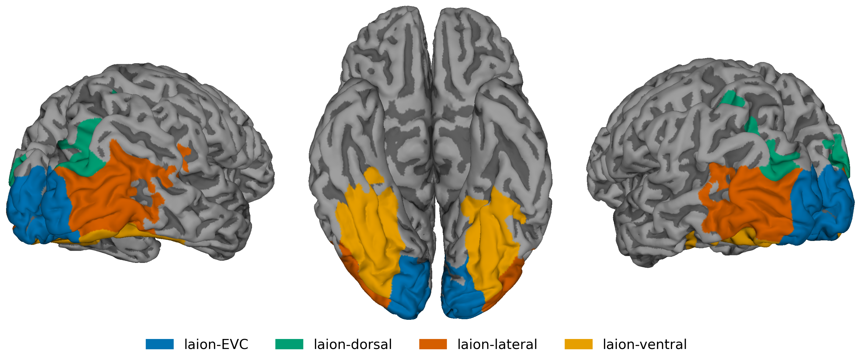
laion-general and its dorsal / lateral / ventral subdivisions.
laion-general covers all cortical regions showing visual responses in the LAION experiment (GLMsingle R²), including isolated patches in IPS and pSTS. laion-EVC comprises the intersection of the V1–V3 with the laion-general selectivity mask.
Note
laion-EVC is not to be confused with the retinotopic EVC mask (union of V1–V3). laion-EVC is restricted to vertices showing significant responses in the LAION experiment and is typically smaller than the retinotopically defined EVC.
The remainder of laion-general is subdivided into three mutually exclusive anatomical parcels: laion-dorsal, laion-lateral, and laion-ventral. These parcels reflect broad cortical territories with anatomically drawn borders and are not intended as functional stream definitions.
The following borders are defined via anatomical landmarks and retinotopy.
Posterior boundary (shared with EVC): the outer border of V3 as defined by the retinotopic delineation.
Dorsal/lateral boundary: runs roughly along the middle temporal gyrus (MTG), tracing the gap between retinotopic areas LO and V3A/V3B.
Lateral/ventral boundary: runs along the inferior occipital–inferior temporal gyrus (IOG–ITG).
This yields three broad visual sectors:
laion-dorsal - cortex dorsal to the LO–V3A/B boundary, covering dorsal occipital and parietal regions.
laion-lateral - cortex between the MTG boundary (dorsal) and the IOG–ITG boundary (ventral), covering lateral occipitotemporal cortex.
laion-ventral - cortex ventral to the IOG–ITG boundary, covering the ventral occipitotemporal surface.
Available Spaces
Per-subject ROIs are released in two spaces:
``fsnative`` - surface masks on each subject’s FreeSurfer surface, as
.func.gii(one value per vertex) and.labelfiles, per hemisphere.``T1w`` (1.8 mm) - volumetric masks resampled to the subject’s T1w grid at the functional resolution, as
.nii.gz.
Both spaces cover the same ROI sets. Surface masks are the canonical reference; the T1w volumes are derived from them and provided for convenience in volume-based analyses.
References
Gao JS, Huth AG, Lescroart MD and Gallant JL (2015) Pycortex: an interactive surface visualizer for fMRI. Front. Neuroinform. 9:23. doi: 10.3389/fninf.2015.00023